Pathology Outlines Herpes simplex esophagitis
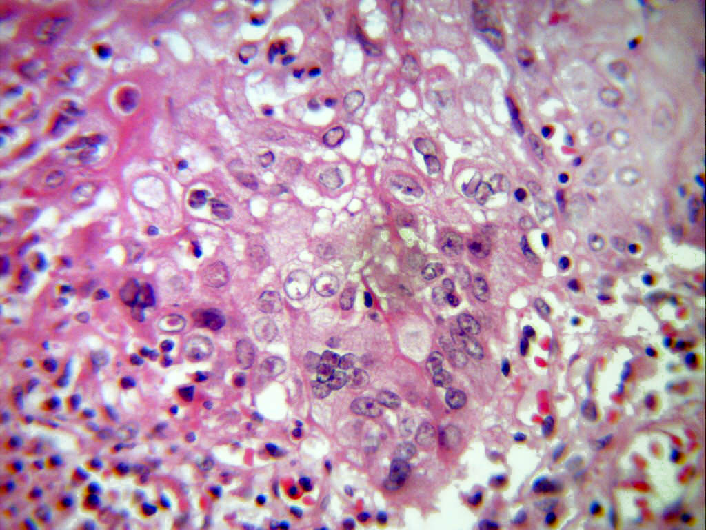
Disseminated herpes infection is commonly seen in immunocompromised patients (vesicular eruptions, encephalitis, esophagitis, keratitis). In the liver there are macroscopic yellow foci of necrosis and hemorrhage. Groups of hepatocytes surrounding these areas contain intranuclear eosinophilic viral bodies (Cowdry type A inclusions).

Histological features of Herpes Simplex Esophagitis showing numerous... Download Scientific
Histopathologically, necrobiotic tubular cells are classified into inclusion-bearing cells of three types: 1) "smudge cells," 2) "Cowdry A" intranuclear inclusion cells including intranuclear.
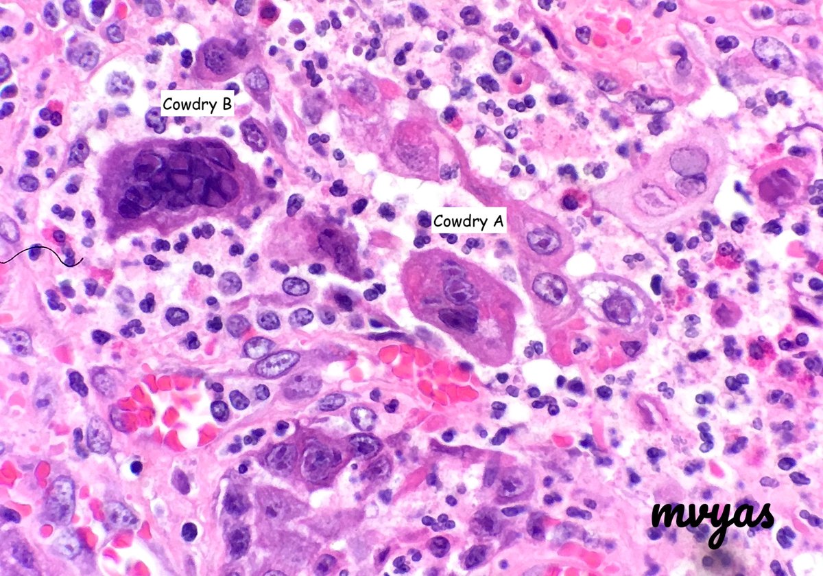
Monika Vyas on Twitter "Nice example of Cowdry A & B inclusions in Herpes esophagitis. Cowdry A
Type A: acidophilic material of droplet-like masses surrounded by clear halos within nuclei., seen in gingivostomatitis and conjunctivitis caused by Herpes simplex virus and also chicken pox caused by varicella zoster. 3 Type B intranuclear eosinophilic without any nuclear change, seen in infection with poliovirus and CMV. History.

Cowdry type A viral inclusions in type 2 pneumocytes (H&E, × 1000, oil... Download Scientific
1 Viral inclusions - types Cowdry types: Cowdry type A inclusion: [2] Round eosinophilic material surrounded by a clear halo. Cowdry type B inclusion: [3] Neuropathology thingy. (???) Images: Cowdry A inclusion (daff.gov.au). Cowdry type A & type B inclusions (altervista.org). Viruses Herpes simplex virus

Ultrastructure of Cowdry type A inclusions Semantic Scholar
Cowdry bodies are eosinophilic or basophilic [1] nuclear inclusions composed of nucleic acid and protein seen in cells infected with Herpes simplex virus, Varicella-zoster virus, and Cytomegalovirus. They are named after Edmund Cowdry. There are two types of intranuclear Cowdry bodies: Type A (as seen in herpes simplex and VZV) [2]

The gill of P. indicus infected with WSSV and Cowdry type A in the... Download Scientific Diagram
Cow·dry type A in·clu·sion bo·dies ( kow'drē tīp in-klū'zhŭn bod'ēz) Dropletlike masses of acidophilic material surrounded by clear halos within nuclei, with margination of chromatin on the nuclear membrane as seen in human herpesvirus-infected cells. Medical Dictionary for the Health Professions and Nursing © Farlex 2012 Cowdry,
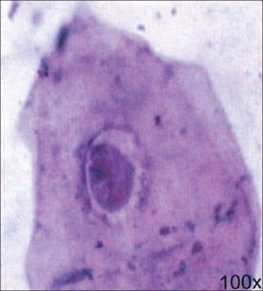
Photomicrograph showing PAPstained, Cowdry Type A incl Openi
OHL was identified in 2 cases (1.67%). In both, the three EBV induced nuclear alterations were observed: Cowdry type A inclusions (Figure 1), ground-glass nuclei (Figure 2) and nuclear beading.

Photomicrograph showing several large Cowdry Type A inclusion bodies.... Download High
https://usmleqa.com/http://usmlefasttrack.com/?p=5369 Lab, Findings:, Cowdry, Type, A, Bodies, (HSV, or, CMV),, Ferruginous, bodies, &, Squamous, Cell, Carc.
Histological features of herpes esophagitis showing Cowdry type A... Download Scientific Diagram
(A) Cowdry type B inclusion of Herpes simplex virus (HSV). There is multinucleation in this infected cell, molding of these nuclei, and chromatin margination beneath the nuclear membrane (Papanicolaou stain, 400×). (B) Cowdry type A inclusion of HSV. Note the characteristic eosinophilic intranuclear inclusion surrounded by a clear zone in.

The three types of inclusion bodies Cowdry A(long arrow), fulltype... Download Scientific
When present in herpes virus infection and present with the other nuclear changes of this infection they are called Cowdry Type A inclusions. Cowdry Type B inclusions are associated with other infections such as poliovirus and do not have the other nuclear changes of herpes infection.

Cowdry type A viral inclusions in type 2 pneumocytes (H&E, × 1000, oil... Download Scientific
Bottom right, a classic Cowdry type A inclusion body is seen in the nucleus of a glial cell at the center of the field (case 10). Note the peripheralizadon of the chromatin at the nuclear membrane and the clear halo surrounding the central eosinophilic, proteinaceous inclusion. (Hematoxylin-eosin stain, x 1,000.) In seven cases, tissue had also.
Histological features of herpes esophagitis showing Cowdry type A... Download Scientific Diagram
Herpes simplex virus infections may be caused by two virus genotypes: herpes simplex virus type 1 and herpes simplex virus type 2 ().Worldwide seroprevalence is high, with antibodies detectable in over 90% of the population. Of these cases, approx. 60% are caused by HSV-1.The most common infections are labial and genital herpes, which present with painful ulcerations.

Ultrastructure of Cowdry type A inclusions Semantic Scholar
Gross description. According to macroscopic appearance, HSV esophagitis is divided into 3 types ( Medicine (Baltimore) 2016;95:e3187 ): Type I: small, punched out lesions with raised margins usually coated with yellowish exudate. Type II: small, punched out lesions but no raised margins or exudate.
The three types of inclusion bodies Cowdry A(long arrow), fulltype... Download Scientific
Here, we report Cowdry type A inclusion bodies (CAIB) in the pancreas as a diagnostic histopathological feature found in adult Nile tilapia naturally infected with TiPV. This type of inclusion body has been well-known as a histopathological landmark for the diagnosis of other parvoviral infections in shrimp and terrestrial species.
Protothaca staminea and Crassadoma gigantea. Histological sections and... Download Scientific
From the Department of Pathology, Regional Primate Research Center, and the Department of Pediatrics, University of Wisconsin School of Medicine, and the John A. Hartford Research Laboratory, Madison General Hospital, Madison, Wis.

Histopathology of IHHNV infected adult P. monodon. Note the Cowdry type... Download Scientific
There are two types: Type A (in herpes infection and yellow fever) and Type-B (in infection with polio and adenovirus) Cowdry type-A inclusion bodies appear as droplet-like masses of acidophilic materials surrounded by clear halos within nuclei, with margination of chromatin on the nuclear membrane. Type-B bodies are not associated with any.